

What You Should Look for When Buying an In Vivo Imaging System
Whether you are looking to add in vivo imaging capabilities to your preclinical workflow, or to upgrade an older system to improve your current labs capabilities, here are some key features to look for while evaluating different options.
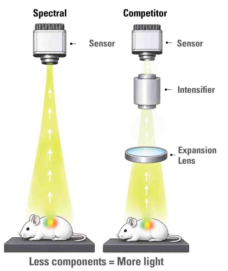
Look for Simplicity in the Optical Path
Additional optical components collectively reduce the efficiency of the system. Every additional mirror, lens, and filter reduces the excitation or detectable photons by several percent, requiring higher illumination power or longer exposure times to compensate. Just two or three additional optical components used to correct non-efficiencies in the light path can reduce the signal by 20% or beyond.
Common optical components to note include:
- Expander lenses – used to expand the field of view to match emitted light to the detector size
- Fiber optic cables or liquid light guides used in the excitation path of some older illumination technologies
- Excitation and emission filters used to reduce non-specific light from broad, white light sources.
Additional optical components also increase the surface area that can collect dust over time and require cleaning. Dust results in light scattering, reduced transmission, and may even produce artifacts in the final image data, skewing quantification. Minimizing the number of optical components and ensuring a closed light path reduces these risks.
Minimum Detectable Radiance (MDR): The Gold Standard for Quantifying System Sensitivity
Sensitivity is a vital performance metric for in vivo imaging systems intended to image faint signals in deep tissue – therefore, it is necessary to understand how minimum detectable radiance (MDR) is calculated and how to properly use it to compare sensitivity of different systems. MDR is defined as the radiance (photons/sec/cm2/sr) required to produce a signal-to-noise ratio of 1; the lowest signal a system can detect. When properly used, MDR provides a means for objective comparison between optical instruments.
MDR is the collective measure of a system’s effective sensitivity with all physical optical components and imaging settings taken into account. Therefore, you must fully understand the parameters used to calculate reported MDR values to ensure objective comparisons between systems. Differences in parameters such as exposure time or pixel binning will affect the resultant MDR and can skew results. When system settings are comparable, MDR is the simplest and most objective means of comparing systems.
Other methods to measure and compare system sensitivity are sometimes reported, including signal-to-background ratio (SBR) or minimum number of detectable cells. However, these sensitivity measurements must be derived from biological samples, which are inherently variable and only add complexity to the question being asked. This is where the frequently cited SBR or “single-cell” sensitivity metrics are quite misleading. “Single-Cell” detection is primarily dependent on the depth of the cell and the reporter used. The ability to detect a single cell or increase SBR is primarily a function of the biological side of the equation, less-so the system itself. For example, a single cell may be detectable if injected subcutaneously where there is minimal attenuation from tissue, but this same cell would become quickly undetectable at greater depth. The same is true for SBR; the greater the depth, the lower the SBR. Each of these metrics is also affected by the reporter used in terms of its relative “brightness” and emitted wavelength, as red-shifted and near infrared (NIR) reporters are detectable at greater depth than reporters in the green and blue regions. For evaluating the actual system hardware capabilities, use of a stable source (phantom) is strongly recommended for objective, comparable, repeatable measures a system’s MDR.
Look for a Well-Controlled and Stable Dark Current and Low Noise Floor
With all other parameters being equal, a lower noise floor indicates a system which can record lower signals and is therefore more sensitive. Lower noise floor means you can detect lower numbers of cells, earlier time points, and signal that is deeper in tissue.
For faint signal detection requiring long exposures, CCDs are the preferred detector technology over electron multiplying CCDs (EMCCD) and sCMOS detectors due to preferential noise properties. EMCCD sensors are extremely sensitive for shorter exposure times with low read noise, but accumulate and amplify dark currents during longer exposures. sCMOS sensors similarly have advantages at short exposure times, but have significantly higher dark currents, and structured non-uniform noise across the sensors. This results in a ‘glow’ artifact that appears with long exposures.
For in vivo imaging systems, the determinant factor of noise is dark current generated by the camera. Dark current is exponentially related to the temperature of the sensor. Dark current approximately halves with every 6-7°C reduction in temperature of the CCD. Therefore, camera cooling is the most critical element for reducing the noise floor and increasing the effective sensitivity of a CCD-based in vivo imaging system. However, not all cooling techniques are equally effective or appropriate for in vivo imaging systems.
The most common sensor cooling options are liquid or air (Peltier) cooling systems. Cryogen-free air cooling is the most modern and preferred method due to their ease of use and component stability. Air cooled systems can effectively cool the sensor down to absolute -90°C, without the potential risk of fluid leaks which can happen with legacy liquid cooling systems. Quick and stable detector cooling is essential for reliable, efficient, and repeatable imaging systems. Changes in the sensor temperature (i.e., delta cooling) over time results in noise properties that are also inconsistent and will thereby impact the quality and reproducibility of signal.
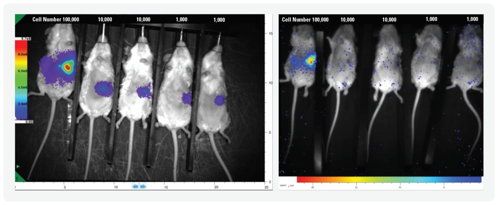
FIGURE LEGEND: Images courtesy of Dr. Lacey McNally, The lefthand image shows data from the AMI HTX with cooling at -90°C Absolute, vs -20°C cooling from a competitor’s systems on the right. Superior cooling reduces dark current and improves low signal detection.
Look for a System Designed for Overall Stability
Often overlooked and underappreciated, stability is among the most defining factors when assessing the quality of an imaging instrument. Strong system stability means consistent, reproducible data from experiments and ultimately better statistical outcomes.
Stability, in a quantifiable sense, is the sum of your system’s noise properties. These noise properties are affected by any component which changes or degrades over time. Ultimately, this has an impact on the consistency with which the system measures photons. For example, older systems may use halogen or mercury halide lamps for illumination. Mercury halide lamps are known to have diminished light intensity as they age, as well as shifts in their spectral output over time. Over the course of their lifetime, illumination profiles change measurably, which also results in differences in experimental data. Mercury halide lamps can also be influenced by strong magnetic or electrical fields, even when they reach a stable operating condition. Bulb-based illumination sources should therefore be avoided in favor of more modern and stable alternatives such as LEDs.
LEDs are stable in both intensity and spectrum over their lifetime and do not require warming up (unlike mercury halide lamps). LEDs also have a narrow spectral profile, thereby requiring minimal filtering within the optical pathway. White light illumination from mercury halide bulbs requires significant intervention from excitation and emission filters to prevent out-of-band excitation light from becoming a source of noise (sometimes inaccurately referred to as “autofluorescence”). The intense light of mercury halide lamps also significantly degrades these filters over time, resulting in lesser light transmission and spectral band shifting. No filter is perfect, so even under optimal conditions, some out-of-band photons will transmit through the filters and be detected, adding system-derived background noise to the resultant images. This is especially pervasive with older systems due to the degradation of the mercury halide bulbs and filters in the light path.
Short wavelength LEDs do not require significant filtering at the excitation side, as they do not produce photons outside of their narrowly-defined range. This results in an illumination system which is stable over the lifetime of the instrument, and therefore, across studies spanning many years. The narrow bandwidth illumination from LEDs results in greater specificity of excitation for fluorescent reporters, as only specified wavelengths are penetrating the tissue and exciting the target fluorophore. This enhanced specificity further allows for native multiplexing of fluorescent reporters in vivo – without the need for subtractive methods such as spectral unmixing.

Look for a Calibration Standard Rigorous Enough for Scientific Studies
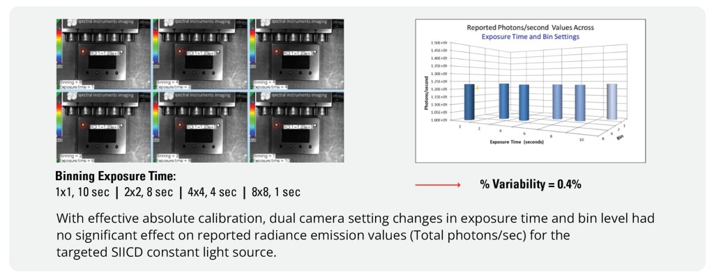
Users should be confident that images taken on different days with different imaging parameters are directly comparable and suitable for publication. Well-designed systems output absolute-calibrated units – values which are normalized for system-derived variables (e.g., camera bias, dark noise) and imaging parameters (e.g., exposure time, binning). For any given light source, absolute calibration should always yield the same value, regardless of when the image was acquired, what camera settings were used or if the source was dim or bright or large or small. Almost by the definition of longitudinal studies, the light sources will dramatically change size and intensity, quality absolute calibration is a must. This process is difficult for many systems, especially those using sCMOS or intensified cameras and even some competitor’s CCD based systems lose data due to imaging parameters like binning. Data taken days, months, or years later is directly comparable to data taken today. Data is directly comparable between different systems across the world.
There’s More Than Just Hard Specs
Engineering specifications define the boundaries of what a system can achieve, but useability is defined by what you, the user, will have to interact with day-to-day. It is worth spending some time to consider how using the system hardware and software fits into your workflow. Likewise, responsive service and support are also important to discuss with prospective vendors: case studies, customer references and understanding field service resources for the lifetime of your system.
With all these factors in mind, you can make an educated evaluation on which systems and features best fit your research.
Reveals Complex Biological Processes
without being overly complicated
-
Easy to Use Software
-
Unmatched Sensitivity
-
High Throughput
-
Free Analysis Software
-
Industry Leading Uptime